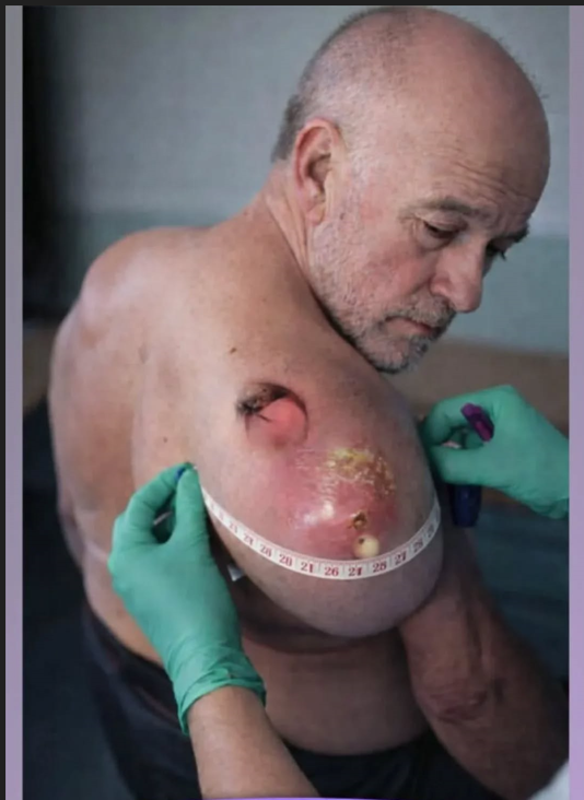
Severe soft tissue lesions of the upper extremity present complex diagnostic and therapeutic challenges. The image depicts a large, erythematous, dome-shaped swelling over the lateral aspect of the upper arm in an elderly male patient. The lesion demonstrates features including significant edema, surface tension, erythema, areas of crusting, ulceration, and possible purulent exudate. The arm circumference is being measured, indicating objective monitoring of inflammatory expansion or mass progression.
Such a presentation warrants immediate clinical attention due to the potential for systemic complications.
✨Clinical Characteristics
The lesion appears markedly inflamed, with surrounding erythema extending beyond the primary swelling. The glossy, stretched skin suggests substantial underlying fluid or tissue expansion. Yellowish crusting may indicate necrosis or purulent drainage. An additional smaller opening superiorly suggests either a sinus tract or secondary ulceration.
In elderly patients, large soft tissue swellings can progress rapidly, especially if underlying immunocompromise, diabetes mellitus, vascular insufficiency, or malignancy is present.
⭐Differential Diagnosis
Several conditions must be considered:
-
Severe Cutaneous Abscess
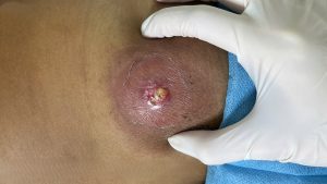
A localized bacterial infection, often due to Staphylococcus aureus, can cause significant swelling and pus accumulation. If untreated, abscesses may enlarge dramatically and form draining sinuses. -
Necrotizing Soft Tissue Infection
Though the image does not definitively show necrotic fascia, rapid expansion, severe erythema, and systemic symptoms would raise concern for necrotizing infection, a surgical emergency. -
Inflamed Epidermoid or Sebaceous Cyst
Long-standing cysts can become infected, resulting in sudden enlargement, tenderness, and purulent drainage. -
Soft Tissue Sarcoma
Large, progressively enlarging masses in older adults must raise suspicion for malignancy. Sarcomas may ulcerate and become secondarily infected. -
Advanced Cutaneous Squamous Cell Carcinoma
Chronic ulcerative lesions with irregular margins and surface breakdown may represent malignant transformation. -
Chronic Osteomyelitis with Soft Tissue Extension
Infections of underlying bone may extend outward, particularly in immunocompromised individuals.
🌙Clinical Assessment
Evaluation should include:
-
Full vital signs to assess systemic inflammatory response.
-
Laboratory testing (CBC, CRP, ESR, blood cultures if febrile).
-
Imaging (ultrasound to assess fluid collection; MRI for deeper involvement).
-
Tissue sampling or biopsy if malignancy is suspected.
-
Microbiological culture of any purulent material.
Measurement of arm circumference, as shown in the image, is useful in tracking progression of edema and inflammatory burden.
💰Management Strategies
Treatment depends on the underlying etiology:
-
Abscess: Incision and drainage, antibiotic therapy guided by culture.
-
Cellulitis: Empiric broad-spectrum antibiotics with adjustment based on response.
-
Necrotizing infection: Immediate surgical debridement.
-
Malignancy: Oncologic referral for biopsy and staging.
-
Chronic infected cyst: Surgical excision after infection control.
Pain management and supportive care are essential. In elderly patients, comorbid conditions must be carefully managed to prevent systemic complications such as sepsis.
⚡Complications
Untreated severe soft tissue lesions may lead to:
-
Sepsis
-
Septic shock
-
Tissue necrosis
-
Limb dysfunction
-
Chronic ulceration
-
Metastasis (if malignant)
Early intervention dramatically improves outcomes.
💨Conclusion
Large, inflamed soft tissue masses in elderly patients require urgent multidisciplinary evaluation. The lesion depicted demonstrates high-risk characteristics including significant swelling, surface ulceration, and inflammatory spread. Accurate diagnosis through imaging and tissue sampling is essential to determine whether the process is infectious, inflammatory, or malignant. Prompt and appropriate management can prevent life-threatening complications and preserve limb function.