What Is That Bump on My Face?
It starts as a small, painless lump under the skin. Then it grows. It might redden, swell, or even develop a small opening at its surface. For many people, a bump that appears near the chin, cheeks, or jawline is dismissed as an aggressive pimple — but what’s seen in the image above tells a very different story.
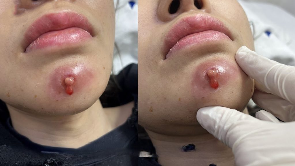
The photographs show a facial skin cyst — specifically what appears to be an epidermal inclusion cyst (also called an epidermoid cyst or sebaceous cyst) that has become inflamed and begun to rupture through the surface of the skin. The left image shows the cyst prior to clinical examination; the right image shows a gloved clinician palpating the area during assessment. This kind of presentation is common in dermatology and cosmetic surgery clinics worldwide.
Types of Skin Cysts That Appear on the Face
Not every bump is the same. The face is home to several distinct types of cysts, each with its own origin, behavior, and preferred treatment approach. Understanding which type you’re dealing with shapes every clinical decision that follows.
Epidermoid Cysts (Sebaceous Cysts)
The most common type. Epidermoid cysts form when surface skin cells migrate into the deeper layers of the dermis and begin to multiply. The cyst wall is made of epithelial tissue, and the interior is packed with keratin — a soft, cheese-like protein that the cells continuously produce. Despite their informal name “sebaceous cysts,” they don’t actually originate from sebaceous glands; that term has largely fallen out of favor in clinical practice.
They can appear anywhere on the face but are especially common near the chin, jaw, cheeks, and behind the ears. When the cyst wall remains intact, they’re typically painless, slow-growing, and mobile under the skin. When they become infected or rupture — as seen in the case above — they cause significant inflammation, redness, and discomfort.
Pilar Cysts
These arise from the outer root sheath of hair follicles and are most often found on the scalp, though they can occasionally appear on the face. Pilar cysts have thicker walls than epidermoid cysts and rarely become infected. Their interior is also keratin-filled, but with a somewhat different composition that gives them a firmer, more rubbery texture on palpation.
Milia
Tiny, white, dome-shaped cysts — typically 1–2 mm in diameter — that develop when dead skin cells become trapped beneath the surface. Unlike epidermoid cysts, milia don’t have a true cyst wall. They’re extremely common in newborns but also appear in adults, especially around the eyes, nose, and cheeks. They’re benign and often resolve on their own, though they can be extracted by a dermatologist if cosmetically bothersome.
Dermoid Cysts
A less common but important category. Dermoid cysts are congenital — they form during fetal development when skin and skin structures become trapped along embryonic fusion lines. On the face, they most commonly appear near the outer eyebrow or at the bridge of the nose. Unlike other cysts, dermoid cysts may contain hair, sweat glands, or even teeth, and they typically require surgical excision.
Clinical Quick Facts
- Epidermoid cysts affect roughly 1 in 100 people
- They are benign — malignant transformation is extremely rare
- Infection occurs in an estimated 20–35% of cases over time
- The chin and jaw are among the most common facial sites
- Complete cyst wall removal is necessary to prevent recurrence
- Draining alone (without excision) has a near-100% recurrence rate
Why Do Facial Cysts Form?
The short answer: the skin’s natural shedding mechanism goes wrong in a very localized way. Normally, dead skin cells rise to the surface and flake off. When something disrupts this process — a blocked follicle, minor trauma, or an inflammatory condition — cells can become trapped and begin building up beneath the surface.
Several factors increase the likelihood of developing facial cysts:
Acne History
This is the single biggest risk factor. Severe or cystic acne damages follicular walls and can create the ideal conditions for an epidermoid cyst to develop. People who have experienced significant teenage or adult acne are far more likely to develop cysts in their twenties, thirties, and beyond, often in the same regions where their acne was concentrated.
Trauma and Minor Injury
A small cut, a popped pimple that was picked aggressively, or even the repeated friction of wearing a face mask can drive surface skin cells downward, seeding a cyst. This mechanism explains why cysts sometimes appear at sites with no obvious acne history.
Hormonal Fluctuations
The sebaceous glands are exquisitely sensitive to androgens. Testosterone and related hormones stimulate oil production, which can contribute to follicular blockage. Women may notice that existing cysts enlarge around menstruation, and new ones may develop during hormonal transitions like pregnancy or perimenopause.
Genetics
Some families show a strong tendency toward cyst formation. Rare inherited conditions such as Gardner syndrome and Gorlin syndrome are associated with multiple cysts, though isolated cysts in otherwise healthy individuals are far more common and carry no sinister implication.
When Should You See a Doctor?
Many small, stable cysts can be safely monitored without intervention. However, certain signs warrant prompt professional evaluation — and the case photographed above represents one of the clearest examples of when to seek care urgently.
⚠ See a Dermatologist If You Notice:
Rapid growth over days or weeks · Significant pain or throbbing · A warm, red, swollen cyst (signs of infection or abscess) · Spontaneous drainage or rupture · A cyst near your eye, nose, or mouth · Any lesion you can’t confidently identify · A cyst that has previously been treated but returned
The photographs show a cyst that has reached a critical stage: it is visibly inflamed, has developed a punctum (a small opening in the skin surface), and exudate — the tell-tale sign of internal pressure and early rupture. At this point, home treatment is not appropriate. Squeezing or attempting to “pop” the cyst risks driving infected material deeper into the tissue, potentially causing cellulitis or abscess formation that requires hospitalization and intravenous antibiotics.
How Doctors Remove Facial Cysts
Treatment choice depends on the cyst’s size, location, infection status, and the patient’s goals. The three main approaches are outlined below.
1. Incision and Drainage (I&D)
When a cyst is acutely infected or painful, the first priority is decompression. The doctor makes a small incision, expresses the contents, and irrigates the cavity. This provides rapid relief but is explicitly not a cure — the cyst wall remains, and recurrence is virtually certain without subsequent excision. I&D is therefore considered a temporizing measure, not a definitive treatment.
2. Intralesional Steroid Injection
For mildly inflamed cysts that haven’t yet progressed to frank infection, an injection of corticosteroid (usually triamcinolone acetonide) directly into the cyst can dramatically reduce inflammation within 48–72 hours. This is particularly useful for reducing a cyst quickly before a more formal surgical procedure, or for patients who cannot yet commit to excision.
3. Surgical Excision
The gold standard. Under local anesthetic, the surgeon makes a small elliptical incision, carefully dissects the cyst away from surrounding tissue, and removes the entire cyst wall intact. The integrity of this step is critical: any remnant of the sac left behind will regenerate the cyst. A small incision is closed with fine sutures, typically removed within a week. Scarring is usually minimal and continues to fade over the following months.
What to Expect: Step by Step
- ConsultationThe dermatologist or surgeon examines the cyst, takes a history, and may order an ultrasound for larger or deeper lesions to map its boundaries before operating.
- Local AnesthesiaLidocaine (with or without epinephrine) is injected around the cyst to numb the area completely. This is the most uncomfortable part of the procedure — the surgery itself is pain-free.
- Incision & DissectionA scalpel makes a small incision directly over the cyst. Using blunt dissection with scissors or a hemostat, the surgeon separates the cyst wall from the surrounding tissue, being careful not to puncture the sac.
- RemovalThe intact cyst is removed and sent to pathology for routine analysis to confirm its benign nature. If the cyst ruptures during removal, the surgeon meticulously removes all wall fragments.
- Closure & AftercareThe wound is irrigated, closed in layers if deep, and dressed. Patients leave with antibiotic ointment and wound care instructions. Sutures are removed at 5–7 days for facial sites.
Recovery and Aftercare
Facial cyst excision is performed as an outpatient procedure, and most patients return to normal activities the same or following day. The wound site will be tender for a few days and may show some bruising. Key aftercare principles include:
Keep it clean and dry for the first 24 hours. After that, gentle cleaning with mild soap is recommended. Avoid sun exposure to the healing scar, as UV radiation can cause hyperpigmentation in healing tissue. Use SPF 30+ daily once sutures are out. Don’t pick or press on the area — even once healed externally, the deeper tissue continues to repair itself for several months.
Scarring after professional facial cyst excision is generally minor — a faint linear mark that fades significantly over 6–12 months. Silicone gel or sheeting can be used from 4 weeks post-surgery to further minimize scarring if desired. In contrast, cysts that are repeatedly squeezed, infected, or allowed to spontaneously rupture tend to leave larger, more irregular scars due to the prolonged inflammation and tissue damage involved.
Can You Prevent Facial Cysts?
Complete prevention isn’t possible — especially if you’re genetically predisposed. However, certain habits can meaningfully reduce your risk and catch new cysts early:
Treat acne proactively. Don’t let severe inflammatory acne run unchecked. Effective treatment with retinoids, antibiotics, or isotretinoin not only clears existing lesions but reduces the follicular damage that seeds future cysts. Resist the urge to squeeze. Every aggressive extraction is a potential trigger for a future cyst. Protect your skin barrier. A healthy, intact skin barrier is less susceptible to the kind of trauma that initiates cyst formation. Stay informed. Regular skin checks — whether self-performed or by a professional — allow cysts to be identified and treated early, before they enlarge or become infected.