What Exactly Is a Cyst Near the Ear?
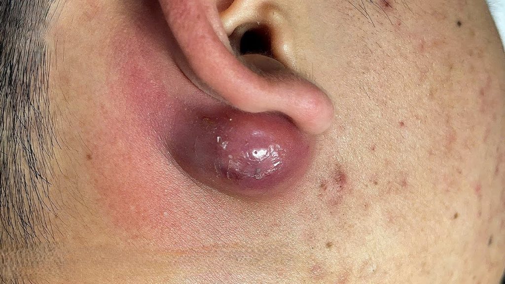
The lump you see in the photograph is a textbook example of an infected epidermoid cyst — sometimes incorrectly called a “sebaceous cyst,” though true sebaceous cysts are actually quite rare. Epidermoid cysts are the most common benign skin lumps in adults, and the area just below the ear lobe, along the jawline, and behind the ear are among their favorite spots to develop.
An epidermoid cyst forms when skin cells (keratinocytes) migrate beneath the surface of the skin rather than shedding normally. The body walls them off in a sac made of stratified squamous epithelium — essentially the same tissue as the outer skin layer. Inside, the sac fills with keratin: a soft, white-to-yellow cheesy material that has a characteristic foul odor when expressed.
On their own, these cysts are harmless and slow-growing. The trouble starts when they become infected — typically by skin-resident bacteria such as Staphylococcus aureus — transforming a benign lump into a painful, red, swollen abscess like the one pictured above.
Why Do They Form Near the Ear?
The periauricular region (the skin surrounding the ear) is particularly prone to cyst formation for several anatomical reasons:
- High follicle density. The skin around the ear contains many hair follicles, which are the most common entry point for inverted skin cells that go on to form cysts.
- Earring puncture sites. Piercing creates a micro-wound that can seed keratinocytes beneath the dermis. Epidermoid cysts at earring holes are extremely common, particularly in people who wear heavy earrings or who experienced an infected piercing.
- Trauma and acne scarring. Past acne lesions, scratches, or surgical scars near the ear can disrupt the normal shedding pathway of skin cells.
- Gardner’s syndrome. In rare cases, multiple epidermoid cysts — particularly around the jaw and ear — may signal this genetic condition, which carries a risk of colorectal cancer. Multiple or recurrent cysts warrant a genetic evaluation.
Stages of an Infected Cyst: From Lump to Abscess
Understanding how a cyst progresses helps explain why the lesion in the photo looks the way it does. Infected epidermoid cysts typically evolve through several stages:
- Quiescent cyst. A small, firm, mobile, skin-colored dome beneath the skin. Usually painless. May have a central punctum (a small dark pore-like opening).
- Early inflammation. Bacteria enter, often after the person squeezes the cyst or it is traumatized. The area becomes mildly tender, pink, and slightly warm.
- Active infection / cellulitis phase. Redness spreads, swelling increases, and the area becomes hot and quite tender. The cyst wall may still be intact.
- Abscess formation. Pus accumulates inside and around the sac. The lesion becomes fluctuant (the center feels soft and “squeezable”). This is the stage depicted in the photograph.
- Spontaneous rupture or resolution. Without treatment, a large cyst may rupture spontaneously, draining purulent material. This brings temporary relief but rarely resolves the underlying sac, leading to recurrence.
Symptoms to Watch For
Local symptoms
- A firm, round lump beneath the skin near the ear
- Pain or tenderness, especially when touched or pressed
- Warmth and redness radiating outward from the center
- Swelling that grows over days
- A soft, “squishy” center (fluctuance) when the abscess forms
- Possible whitish or yellowish discharge if the cyst ruptures
Systemic warning signs — seek immediate care
⚠ Go to an emergency department if you experience:
- Fever above 38.5 °C (101.3 °F) with a rapidly spreading rash
- Red streaks extending from the cyst (signs of lymphangitis)
- Swollen, tender lymph nodes in the neck
- Difficulty opening the jaw or swallowing
- Severe facial swelling affecting the eye or neck
- Feeling generally unwell, confused, or very tired
These may indicate a spreading deep-tissue infection, sepsis, or Ludwig’s angina — life-threatening conditions that require hospital admission.
How Doctors Diagnose It
In most cases, diagnosis is clinical — meaning a trained physician can identify an infected epidermoid cyst by looking at and palpating (examining by touch) the lump. No tests are usually required for a straightforward case.
However, the doctor may order further investigation if:
- The lesion is unusually hard, rapidly growing, or fixed to underlying tissue (to rule out a parotid gland tumor or malignancy)
- There is significant surrounding cellulitis — an ultrasound can confirm the depth and extent of the abscess
- The patient is immunocompromised or diabetic (cultures may be taken to identify the causative bacteria and guide antibiotic selection)
- The cyst recurs multiple times in the same location
A point-of-care ultrasound, increasingly used in emergency and dermatology settings, can also differentiate a cyst from a lymph node, parotid lesion, or vascular structure — all of which can mimic an infected cyst near the ear.
Treatment Options
This is where patient expectations and medical reality often diverge. An infected cyst of the size and severity pictured above cannot be resolved with creams, warm compresses alone, or antibiotics alone. The definitive treatment depends on the stage.
Incision and drainage (I&D)
For a fluctuant abscess like the one in the photograph, incision and drainage is the first-line treatment. A physician makes a small incision into the most fluctuant part of the lesion under local anesthesia, releases the purulent material, and may loosely pack the cavity with gauze to allow continued drainage. Relief is often dramatic and immediate. This procedure does not remove the cyst sac.
Antibiotics
Oral antibiotics (typically a 5–7 day course of a penicillinase-resistant penicillin, a first-generation cephalosporin, or — for MRSA coverage — trimethoprim-sulfamethoxazole or doxycycline) are prescribed when:
- There is significant surrounding cellulitis
- The patient is immunocompromised, diabetic, or has a prosthetic implant
- Systemic signs of infection (fever, chills) are present
Antibiotics alone without drainage are generally insufficient for a frank abscess and should not be relied upon as the sole treatment.
Complete surgical excision
Once the acute infection has settled — usually 4 to 6 weeks after incision and drainage — the definitive cure is complete surgical excision of the cyst sac. The entire sac must be removed intact; if the sac wall is left behind, the cyst will almost certainly recur. This is typically performed under local anesthesia in an outpatient or day-surgery setting and takes 15–30 minutes.
What Not to Do: The Dangers of Squeezing
The impulse to squeeze a large, tense cyst is understandable — especially given how satisfying it looks in the viral “Dr. Pimple Popper” videos that have made dermatology so popular online. However, squeezing an infected cyst near the ear carries real risks:
- Rupture into deeper tissue. Forceful squeezing can rupture the sac wall into the dermis or deeper subcutaneous fat, spreading keratin debris and bacteria far more widely than the original infection occupied. This triggers a severe foreign-body inflammatory reaction that is painful and much harder to treat.
- Seeding nearby structures. Near the ear, this includes the parotid gland, the facial nerve, and the lymph nodes of the neck — none of which you want infected.
- Scarring and incomplete drainage. The thick keratin contents of an epidermoid cyst do not drain efficiently through a pinhole opening. You are unlikely to empty the sac and highly likely to traumatize the overlying skin, leading to scarring.
- Recurrence. Even if the contents partly drain, the sac remains, and the cyst will refill.
Recovery and What to Expect After Treatment
After incision and drainage, most patients experience significant pain relief within 24 hours. The wound typically heals over 1–2 weeks with daily dressing changes. A small amount of continued drainage is expected and normal during this period.
After complete surgical excision:
- Swelling and bruising around the ear and jaw are normal for 3–5 days
- Sutures (if non-absorbable) are removed at 7–10 days
- The area should be kept dry for 48 hours post-operatively
- Sun protection of the scar is important for the first 6–12 months
- The cure rate with complete excision exceeds 95% — recurrence usually indicates residual sac wall
Prevention: Can You Stop Cysts From Forming?
Unfortunately, most epidermoid cysts are not preventable — they arise from normal skin cells behaving abnormally, often without a clear trigger. That said, the following habits may reduce the risk of recurrence or new cyst formation:
- Avoid picking at or squeezing any skin lesion near the ear, as trauma is a known precipitant
- Keep earring sites clean and avoid heavy earrings that cause micro-tears at the piercing channel
- Treat acne early and effectively to minimize skin trauma and scarring
- After successful excision, report any recurrence promptly — early re-excision is simpler than treating a recurrent large cyst
When to See a Doctor: A Summary
If the lump near your ear resembles what is shown in the photograph above — significantly swollen, red, warm, and tender — you should see a doctor within 24–48 hours rather than waiting. There is no safe home treatment for an abscess of this size and severity. Prompt professional drainage not only resolves the infection faster but also prevents the serious complications described above.
If you have a smaller, painless, skin-colored lump near the ear that has been there for months without change, you can arrange a routine dermatology appointment. Elective excision before a cyst becomes infected is always easier, less painful, and more likely to produce a clean cosmetic result.