In the world of dermatology and minor surgery, few things capture the public’s attention like a massive skin anomaly. The image above—showing a densely packed cluster embedded in the skin—combined with the concept of a “gigantic cyst excision,” presents a fascinating look into how complex skin conditions are managed, treated, and closed.
While the internet often labels images like this as “cysts,” a closer look reveals a complex story of diagnosis, removal, and the art of surgical closure.
1. Visual Analysis: Cyst vs. Infestation
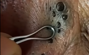
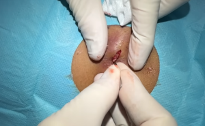
The first thing a surgeon must do is diagnose the lesion. The caption describes a “Gigantic Cyst,” but the visual evidence in the photo presents a pattern known as Trypophobia (fear of clustered holes).
-
The Cyst Theory: A true sebaceous or epidermoid cyst is usually a single, smooth lump under the skin filled with keratin (cheese-like substance). When excised, it leaves a large “dead space” or cavity.
-
The Image Reality: The photo actually depicts what looks like Cutaneous Myiasis (a fly larvae infestation) or a Ticks Cluster. However, seasoned medical observers often identify this specific viral image as a work of Special Effects (SFX) Makeup using beans or seeds and wax to simulate a severe infection.
Regardless of whether the wound is caused by removing a massive cyst sac or extracting a cluster of foreign bodies, the result is the same: A Deep Pocket Defect.
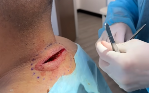
2. The Surgical Challenge: The “Deep Pocket”
Once a mass—be it a cyst sac or a cluster of parasites—is removed, the skin doesn’t just snap back into place. The removal leaves behind a crater known as “Dead Space.”
If a surgeon were to simply stitch the top layer of skin (epidermis) closed over a deep hole, fluids (seroma) or blood (hematoma) would fill the empty space. This creates a breeding ground for bacteria and leads to infection.
This is where the “Closure” mentioned in your text becomes the most critical part of the procedure.
3. The Art of the Closure: Suturing Techniques
As the caption notes (“See how I use sutures to close deep pocket”), closing a wound of this magnitude requires a multi-layered approach. You cannot simply zip it up like a jacket.
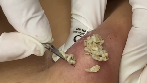
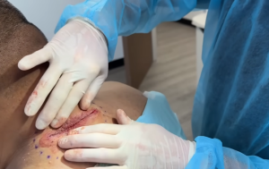
Step A: Undermining
First, the surgeon uses scissors to separate the skin from the underlying tissue around the edges of the hole. This loosens the skin, allowing it to stretch and cover the hole without too much tension.
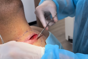
Step B: Deep Buried Sutures
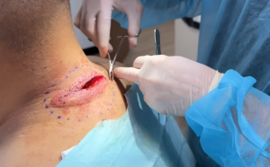
This is the “secret sauce” of closing a giant cyst void.
-
The doctor uses absorbable stitches (sutures that the body dissolves over time).
-
They stitch the bottom and middle layers of the tissue together first.
-
By pulling the deep tissues together, the floor of the hole is raised, and the edges are brought closer. This obliterates the “dead space.”
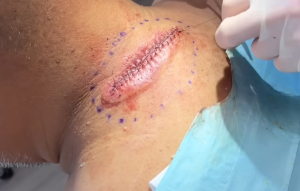
Step C: Superficial Closure
Once the deep pocket is closed internally, the skin edges should lie close together naturally. The final step is using non-absorbable sutures (like nylon) on the surface to create a neat, cosmetic line.
4. Why We Watch: The Psychology of Extraction
Why are videos of cyst excisions and deep pocket closures so popular? It relates to the Grotesque-Relief Cycle.
-
Tension: We see the “monster” (the cyst or cluster) on the body. It looks painful and wrong.
-
Action: The doctor performs the excision. It is messy and chaotic.
-
Resolution: The sutures are placed. The chaotic hole becomes a neat, straight line. Order is restored to the body.
Summary
Whether dealing with a massive cyst removal or the cleanup of a parasitic infection like the one mimicked in the photo, the principles of surgery remain the same. It is not just about “cutting it out”—it is about understanding the anatomy of the skin and knowing how to rebuild the tissue layer by layer so the patient heals without infection or massive scarring.