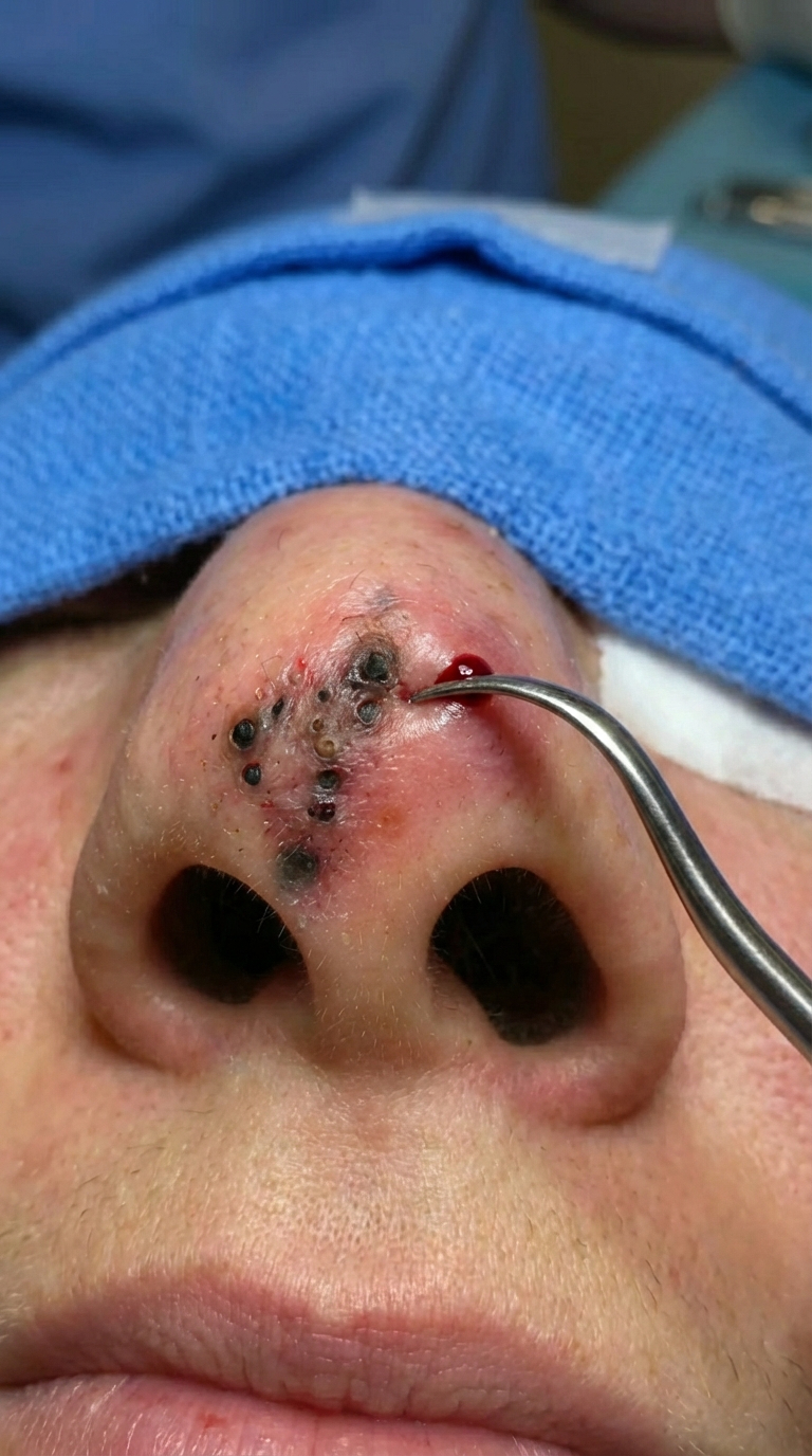
The image above shows a large dark plug inside the nostril opening, surrounded by redness and mild swelling. This presentation is most consistent with a dilated pore filled with oxidized keratin and oil, sometimes referred… Read more
The image above shows a large dark plug inside the nostril opening, surrounded by redness and mild swelling. This presentation is most consistent with a dilated pore filled with oxidized keratin and oil, sometimes referred… Read more
The image above shows a large dark plug inside the nostril opening, surrounded by redness and mild swelling. This presentation is most consistent with a dilated pore filled with oxidized keratin and oil, sometimes referred… Read more
The image above shows a large dark plug inside the nostril opening, surrounded by redness and mild swelling. This presentation is most consistent with a dilated pore filled with oxidized keratin and oil, sometimes referred… Read more
The image above shows a large dark plug inside the nostril opening, surrounded by redness and mild swelling. This presentation is most consistent with a dilated pore filled with oxidized keratin and oil, sometimes referred… Read more
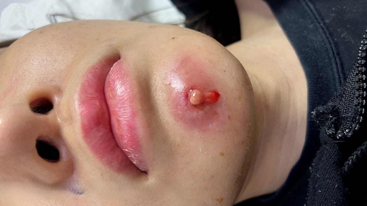
The image above shows a severely inflamed lesion located on the chin, with visible redness, swelling, pus formation, and a dark central core. The surrounding skin appears irritated and thickened, suggesting an advanced infection. Conditions… Read more
The image above shows a severely inflamed lesion located on the chin, with visible redness, swelling, pus formation, and a dark central core. The surrounding skin appears irritated and thickened, suggesting an advanced infection. Conditions… Read more
The image above shows a severely inflamed lesion located on the chin, with visible redness, swelling, pus formation, and a dark central core. The surrounding skin appears irritated and thickened, suggesting an advanced infection. Conditions… Read more
The image above shows a severely inflamed lesion located on the chin, with visible redness, swelling, pus formation, and a dark central core. The surrounding skin appears irritated and thickened, suggesting an advanced infection. Conditions… Read more
The image above shows a severely inflamed lesion located on the chin, with visible redness, swelling, pus formation, and a dark central core. The surrounding skin appears irritated and thickened, suggesting an advanced infection. Conditions… Read more